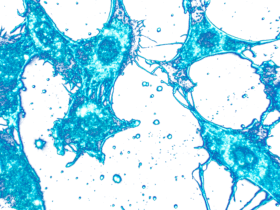
About the Video
The Hidden World of Cell Lines is a short, engaging introduction to cell lines and their problems. It is written for anyone who is new to cell culture and its problems, with no prior knowledge needed. The video takes a fun and quirky approach to the topic, in an effort to capture the viewer’s interest, but the problems it describes are far more serious than the quirky setting may suggest.
Misidentified cell lines
Cell line misidentification is often thought of as a “HeLa problem” – something that only happens if a lab is working with HeLa cells. However, today’s problem with misidentified cell lines is bigger than any one cell line. The ICLAC Register of Misidentified Cell Lines [1] currently lists over 500 cell lines that are known to be misidentified with no known authentic material. The Register lists 154 “contaminating” cell lines; the commonest are HeLa, T-24 and M14.
We chose M14 for our video because it has a fascinating story that clearly shows the impact of cell line misidentification. Scientists have spent years debating the origins of M14 (from a male with melanoma) and MDA-MB-435 (from a female with breast cancer). Authentication testing shows that these two cell lines actually come from the same person [2, 3]. So which one is misidentified?
ICLAC was finally able to put this mystery to rest by testing early samples from the donor of M14. Samples preserved by Dr. Donald Morton – a scientist and clinician who established the cell line in the 1970s – corresponded to M14 and to its original donor’s biological sex and blood type [4].
M14 is authentic, which means that MDA-MB-435 is misidentified and does not come from a female with breast cancer. Using MDA-MB-435 – from a male with melanoma – as a model for breast cancer may lead to inconclusive results, wasted time and resources.
Authentication testing
The key to preventing misidentified cell lines is authentication testing. Authentication uses a genotype-based method to confirm the cell line’s species and compare its genotype to donor material, if available [5]. Suitable methods include short tandem repeat (STR) profiling and single nucleotide polymorphism (SNP) analysis.
STR profiling is the standardized, consensus approach for human cell lines [6]. STR profiling is also available for mouse [7], rat [8, 9], African green monkey [10], and dog [11] cell lines. Search tools are available to compare a lab’s STR profiles to those from cell banks and publications e.g., using the CLASTR search tool available through Cellosaurus [12].
SNP analysis is a complementary approach that is particularly useful for high-throughput, sequence-based platforms [13, 14].
Wasted research time and money
Lack of reproducibility in preclinical research wastes an estimated $56 billion per year in the United States alone [15]. Biological reagents and reference materials are responsible for about a third of that cost. Cell line problems – misidentification, contamination, genetic drift, and clonal evolution – are important causes of irreproducible research and affect everyone who works with cell lines.
Microbial contamination
Any cell line can be contaminated with micro-organisms [16]. Many bacteria, yeasts and fungi cause obvious changes when observed by eye, such as cloudiness of the culture medium or a change in its pH. These micro-organisms may also be visible under the microscope. However, other microbial contaminants are not visible when cell lines are routinely examined under the microscope and need specific testing to be detected. These include mycoplasmas and viruses.
Mycoplasmas (more correctly known as mollicutes) can be distinguished from other bacteria by their small size (0.3-0.8 µm) and lack of a cell wall. These organisms adhere to cultured cells and can invade or fuse with the cell to take up residence within the cytoplasm. They are common contaminants in research laboratories that are not sensitive to widely used antibiotics such as penicillin [17].
All cell lines should be regularly tested for mycoplasma. Various test methods can be used, including PCR [18]. Contaminated cultures should always be discarded unless they are unique and irreplaceable. Procedures are available for treatment of mycoplasma-contaminated cultures [19].
Viral contamination has been detected in 3-5% of human cell lines [20, 21]. Murine leukemia viruses are particularly common, due to xenograft passaging in immunocompromised mice. Testing for viral contaminants may be necessary as part of a safety risk assessment.
Good Cell Culture Practice
Good cell culture practice (GCCP) refers to the principles and practices [22, 23] used to obtain cell lines from reliable sources, handle them safely and with good aseptic technique, maintain stocks at early passage and good quality, and keep records of cell line information. Key cell line information is available using Cellosaurus [24]; always look up a cell line there before you work with it.
Credits
Financial support for the video was provided by the European Tissue Culture Society (ETCS) as a legacy donation. We thank Nathan Burgess and Sid Shukla of The Video and Animation Production Company (THE VAPCO) for the animation, Lint Free Voices for the voiceover, Amanda Capes-Davis for the script and CellBank Australia for assistance with the donation.
References
[1] Capes-Davis A et al. Check your cultures! A list of cross-contaminated or misidentified cell lines. Int J Cancer 2010; 127(1):1-8. doi: 10.1002/ijc.25242.
[2] Rae JM et al. MDA-MB-435 cells are derived from M14 melanoma cells–a loss for breast cancer, but a boon for melanoma research. Breast Cancer Res Treat 2007; 104(1): 13-9. doi: 10.1007/s10549-006-9392-8.
[3] Chambers AF. MDA-MB-435 and M14 cell lines: identical but not M14 melanoma? Cancer Res 2009; 69(13): 5292-3. doi: 10.1158/0008-5472.CAN-09-1528.
[4] Korch C et al. Authentication of M14 melanoma cell line proves misidentification of MDA-MB-435 breast cancer cell line. Int J Cancer 2018; 142(3): 561-572. doi: 10.1002/ijc.31067.
[5] Korch C, Varella-Garcia M. Tackling the human cell line and tissue misidentification problem is needed for reproducible biomedical research. Adv Mol Pathol 2018; 1(1): 209-228.e36. doi: 10.1016/j.yamp.2018.07.003.
[6] ANSI/ATCC ASN-0002-2021 (revised). Human cell line authentication: Standardization of short tandem repeat (STR) profiling.
[7] Almeida JL et al. Interlaboratory study to validate a STR profiling method for intraspecies identification of mouse cell lines. PLoS One 2019; 14(6): e0218412. doi: 10.1371/journal.pone.0218412.
[8] Walder RY et al. Short tandem repeat polymorphic markers for the rat genome from marker-selected libraries. Mamm Genome 1998; 9(12): 1013-21. doi: 10.1007/s003359900917.
[9] Bryda EC, Riley LK. Multiplex microsatellite marker panels for genetic monitoring of common rat strains. J Am Assoc Lab Anim Sci 2008; 47(3): 37-41.
[10] Almeida JL et al. Authentication of African green monkey cell lines using human short tandem repeat markers. BMC Biotechnol 2011; 11: 102. doi: 10.1186/1472-6750-11-102.
[11] O’Donoghue LE et al. Polymerase chain reaction-based species verification and microsatellite analysis for canine cell line validation. J Vet Diagn Invest 2011; 23(4): 780-5. doi: 10.1177/1040638711408064.
[12] Robin T et al. CLASTR: The Cellosaurus STR similarity search tool – A precious help for cell line authentication. Int J Cancer 2020; 146(5): 1299-1306. doi: 10.1002/ijc.32639.
[13] Yu M et al. A resource for cell line authentication, annotation and quality control. Nature 2015; 520(7547): 307-11. doi: 10.1038/nature14397.
[14] Liang-Chu MMY et al. Human biosample authentication using the high-throughput, cost-effective SNPtrace(TM) system. PLoS One 2015; 10(2): e0116218. doi: 10.1371/journal.pone.0116218.
[15] Freedman LP et al. The Economics of Reproducibility in Preclinical Research PLoS Biol 2015; 13(6): e1002165. doi: 10.1371/journal.pbio.1002165.
[16] Fogh J et al. A review of cell culture contaminations. In Vitro 1971; 7(1): 26-41.
[17] Drexler HG, Uphoff CC. Mycoplasma contamination of cell cultures: Incidence, sources, effects, detection, elimination, prevention. Cytotechnology 2002; 39(2): 75-90.
[18] Uphoff CC, Drexler HG Detection of Mycoplasma contamination in cell cultures. Curr. Protoc. Mol. Biol. 2014; 106: 28.4.1–28.4.4. doi: 10.1002/0471142727.mb2804s106.
[19] Uphoff CC, Drexler HG. Eradication of Mycoplasma contaminations from cell cultures. Curr Protoc Mol Biol 2014; 106:28.5.1-12. doi: 10.1002/0471142727.mb2805s106.
[20] Shioda S. et al. Screening for 15 pathogenic viruses in human cell lines registered at the JCRB Cell Bank: characterization of in vitro human cells by viral infection. R Soc Open Sci 2018; 5 (5): 172472. doi: 10.1098/rsos.172472.
[21] Uphoff CC et al. Screening human cell lines for viral infections applying RNA-Seq data analysis. PLoS One 2019; 14 (1): e0210404. doi: 10.1371/journal.pone.0210404.
[22] Coecke S et al. Guidance on good cell culture practice. a report of the second ECVAM task force on good cell culture practice. Altern Lab Anim 2005; 33(3): 261-87. doi: 10.1177/026119290503300313.
[23] Geraghty RJ et al. Guidelines for the use of cell lines in biomedical research. Br J Cancer 2014; 111(6): 1021-46. doi: 10.1038/bjc.2014.166.
[24] Bairoch A. The Cellosaurus, a cell-line knowledge resource. J Biomol Tech 2018; 29(2): 25-38. doi: 10.7171/jbt.18-2902-002.


